Hanyu Frontier
Structural Heart Disease
Frontier Explorer
NEW OPPORTUNITIES FOR INNOVATIVE INVESTMENT IN
STRUCTURAL HEART DISEASE
STRUCTURAL HEART DISEASE

Structural Heart Disease
Frontier Explorer
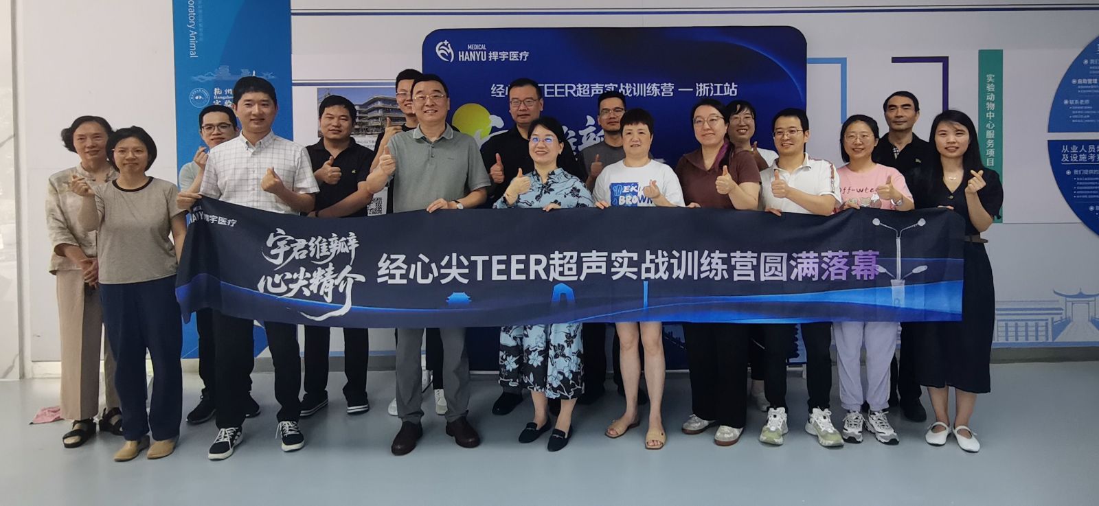
Recently, the 5th session of the “Yujun ValveCare · Transapical Excellence” Transapical TEER Ultrasound Hands-on Training Camp, organized by Hangyu Medical, was successfully held in Hangzhou.
This session focused on ultrasound-guided precision and clinical application in transapical transcatheter edge-to-edge repair (TA-TEER).
The event featured keynote lectures by Professor Dong Lili from the Department of Cardiac Ultrasound, Zhongshan Hospital, Fudan University, and Professor Pan Dihao from the Department of Cardiovascular Surgery, First Affiliated Hospital, Zhejiang University School of Medicine.
Attracting numerous cardiologists and young physicians from across Zhejiang Province, the training camp injected new momentum into the standardized clinical implementation of TA-TEER technology in the region.

Academic Highlights: Bridging Ultrasound Navigation and Clinical Practice
Continuing the dual-track format of “theory + practice,” this session invited leading experts to deconstruct key steps of the TA-TEER workflow, establishing a clear link between imaging cognition and clinical decision-making.
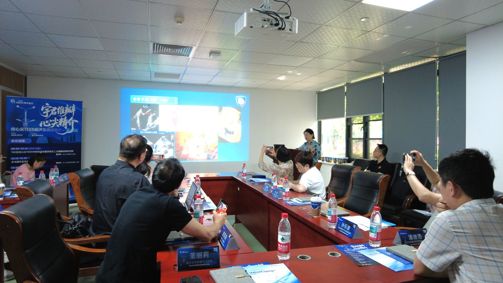 In her lecture, “Intraoperative Ultrasound Views and Anatomic Variation Recognition in TA-TEER”, Prof. Dong emphasized the central role of echocardiography as a surgical navigator.
She introduced the “2+1” intraoperative imaging strategy:
Core view: Mid-esophageal commissural view (50°–70°) for precise identification of A1–A3 and P1–P3 segments.
Complementary view: Long-axis view (120°–135°) for annular alignment.
Auxiliary view: 3D surgical left atrial perspective to overcome challenges in view acquisition and spatial orientation.
She noted that in cases with marked LV enlargement (LVEDD > 70 mm), fine adjustments are crucial to prevent image distortion.
Prof. Dong further clarified ultrasound differentiation between two major types of regurgitation:
DMR (Degenerative MR): Identify prolapse (commonly A2/P2), flail (chordal rupture), and Barlow’s disease (redundant leaflet), and distinguish clefts (discontinuity of the anterior leaflet) from indentations (posterior leaflet folds).
FMR (Functional MR): Differentiate VFMR (ventricular type, papillary muscle displacement) from AFMR (atrial type, annular dilation with “hamstring sign”), stressing that tethering severity predicts procedural success.
She highlighted that in elderly patients, mixed DMR/FMR patterns are common, requiring layer-by-layer evaluation of leaflet thickness, calcification, and jet direction to guide surgical strategy precisely.
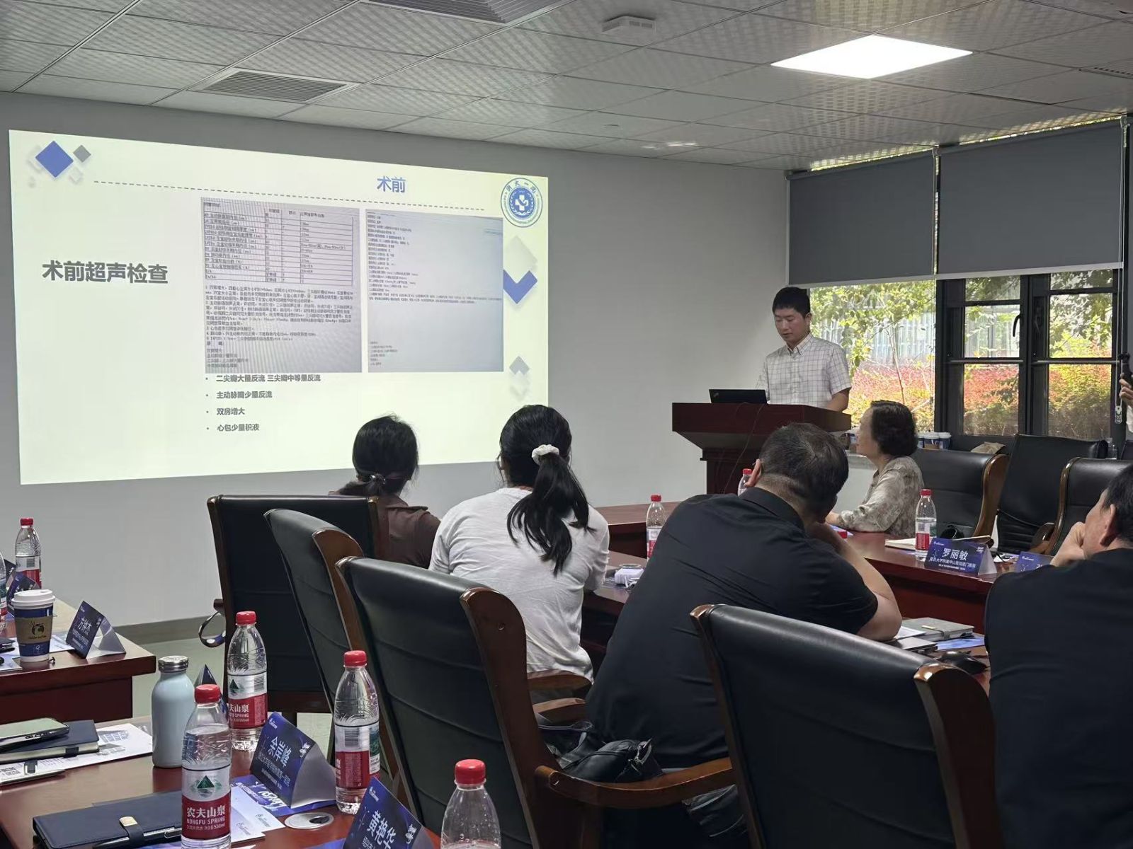
Prof. Pan Dihao shared a recent case of an 82-year-old male with functional MR treated at Zhejiang University First Hospital.
The patient presented with 10 years of atrial fibrillation and biatrial enlargement, with preoperative echo showing severe MR (4+) and multi-jet regurgitation spanning segments 1–3 (width: 20.8 mm).
The core challenge lay in prioritizing jets and optimizing clip orientation.
Key procedural takeaways:
Preoperative planning: Use of MVC-IIIF clip targeting segment 1 jet, with intraoperative assessment guiding the need for a second clip to prevent excessive tension.
Intraoperative technique: A 16F sheath was advanced into the LV via guidewire; under 3D surgical visualization, clip rotation was adjusted to align perpendicular to the coaptation line. The first clip achieved a stable double-orifice morphology, with pure echo guidance (no DSA) throughout.
Postoperative outcomes: Device time was 10 minutes, residual MR reduced to 1+, anterior/posterior leaflet grasping lengths were 8.9 mm / 14.7 mm, and mean transmitral gradient was 1 mmHg.
At 30-day follow-up, MR remained mild-to-moderate with significant functional improvement.
Prof. Pan stressed adherence to the “4M Principles” — grasping length, residual MR, mean gradient, and tissue bridge morphology — as the gold standard for ensuring safe and effective clip release.
During the expert panel, participants discussed surgical strategies for mitral regurgitation — spanning open repair, valve clamping, and replacement techniques.
The conversation delved into lesion characterization, procedural difficulty, and decision-making in complex MR cases, fostering deep clinical exchange between imaging and surgical teams.


The afternoon session transitioned from theory to immersive simulation-based training, where participants completed four structured modules:
TEE Standard View Acquisition: Rapid switching between commissural and long-axis views with stable image control.
Apical Puncture Guidance: Simulated puncture site localization using pre-procedural imaging, mastering the “cotton swab marking” technique to avoid vascular zones.
Clip Performance Evaluation: Real-time tracking of clip position and assessing outcomes via morphology, residual MR, mean gradient, and grasping depth.
Complication Management: Simulated scenarios such as chordal entanglement and leaflet slippage, emphasizing adaptive troubleshooting.
Under expert supervision, trainees practiced hands-on with direct feedback, strengthening eye–hand coordination between ultrasound imaging and procedural execution, truly achieving “learning by practicing.”

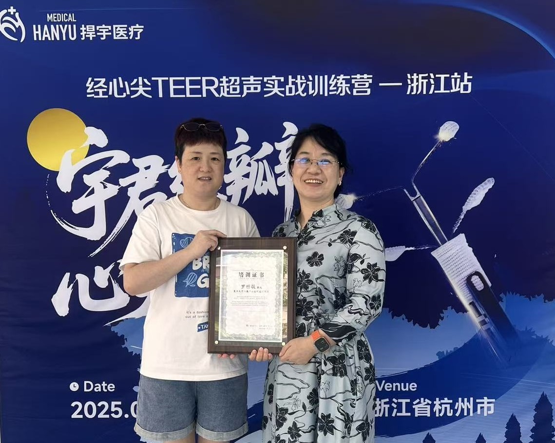
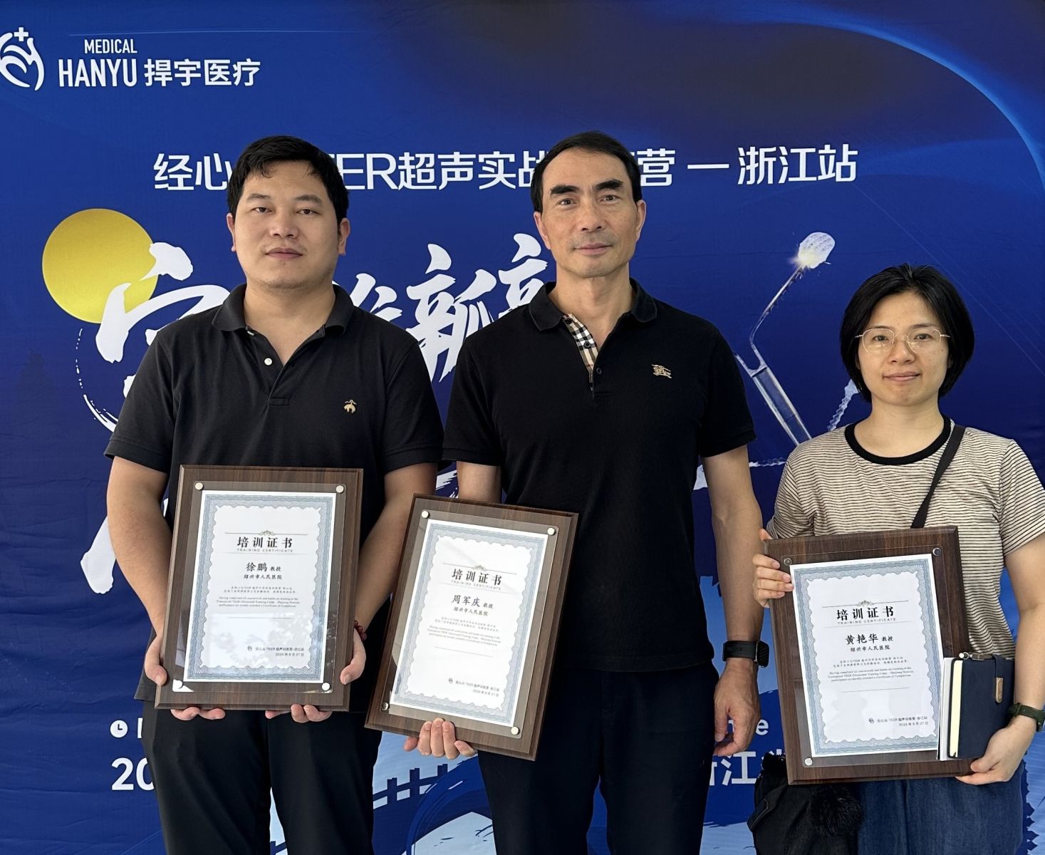
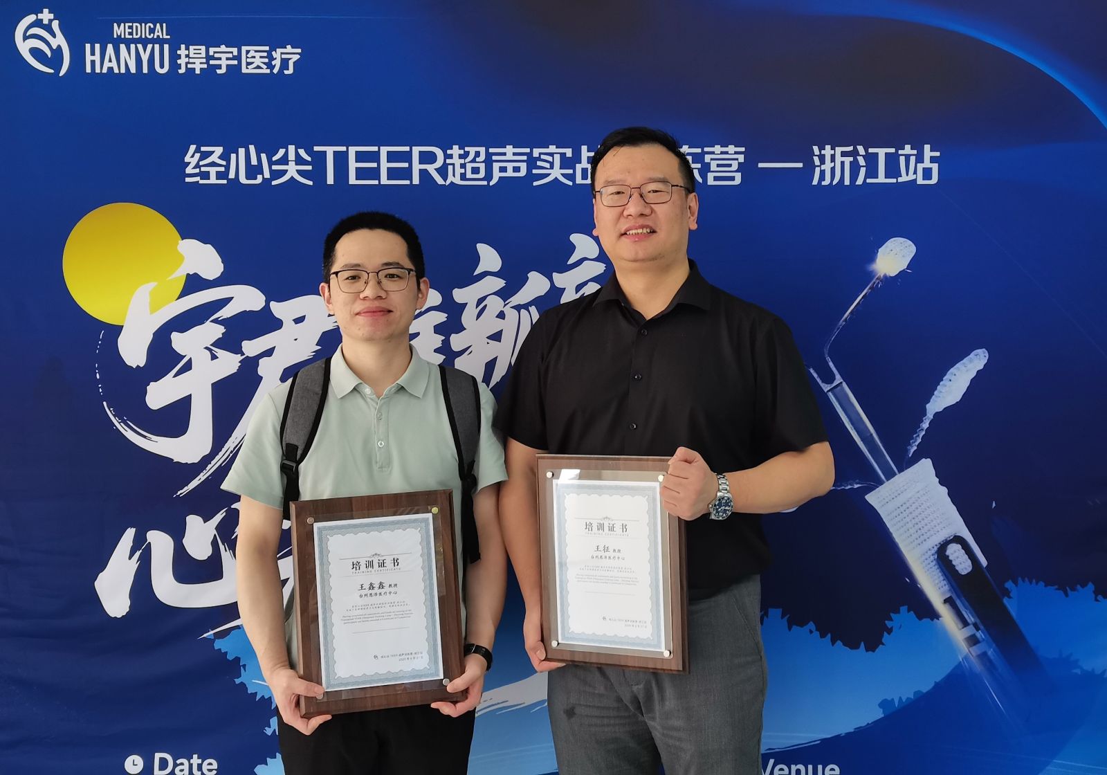
At the conclusion of the camp, participants who completed all modules received certificates of completion, marking their acquisition of fundamental TA-TEER ultrasound-guided procedural competence.
Attendees expressed that the course effectively bridged the gap between ultrasound theory and clinical practice, especially in complex anatomical recognition and multi-jet MR management, providing invaluable guidance for future interventions.
As the core national platform for the dissemination of domestic TA-TEER technology, the “Yujun ValveCare · Transapical Excellence” program will continue to expand across Zhejiang and neighboring regions.
Through the “Expert Mentorship + Practical Training” model, it aims to cultivate more multidisciplinary specialists skilled in both imaging and intervention.
Looking ahead, Hangyu Medical will continue to partner with leading clinical centers nationwide to promote the standardization and precision development of TA-TEER, enabling more elderly and high-risk MR patients to benefit from minimally invasive therapy.


TEL: 021-6219 9996
ADD:Building 14, Gemdale Weixin Technology Park, 1288 Zhongchun Road, Minhang District, Shanghai