Hanyu Frontier
Structural Heart Disease
Frontier Explorer
NEW OPPORTUNITIES FOR INNOVATIVE INVESTMENT IN
STRUCTURAL HEART DISEASE
STRUCTURAL HEART DISEASE

Structural Heart Disease
Frontier Explorer
In July 2025, the TEER Team led by Professor Yan Yang from the Department of Cardiovascular Surgery, First Affiliated Hospital of Xi'an Jiaotong University, collaborated closely with teams from Anesthesiology, Operating Room, and Echocardiography to successfully perform two transcatheter mitral valve repair (TEER) procedures in a single day. One of the cases involved a typical commissural lesion in Zone 3, which was repaired using the domestic mitral valve clip system ValveClamp® Type II. The procedure was completed in a short time: after clipping, regurgitation disappeared completely, and the clip integrated seamlessly with the commissure to form a large single orifice. The intraoperative performance was excellent, and the clipping effect was described as "natural and flawless".
Case Introduction
Preoperative Evaluation
Imaging evaluation revealed that the lesion was located at the Zone 3 commissure of the mitral valve, with complex valve anatomy. The patient had hypoxemia and could not tolerate cardiopulmonary bypass, making traditional surgical repair high-risk. Minimally invasive TEER was therefore selected as the optimal treatment. The surgical team used the ValveClamp® Type II clip for precise positioning and clipping. The procedure was completed quickly and efficiently through an accelerated anesthesia pathway. Under real-time guidance of intraoperative transesophageal echocardiography (TEE), the clip was accurately placed at the Zone 3 commissure, achieving stable leaflet capture and a natural clipping line. Mitral regurgitation was significantly improved. The endotracheal tube was removed immediately after surgery; no postoperative complications occurred, and the patient’s cardiac function recovered well.
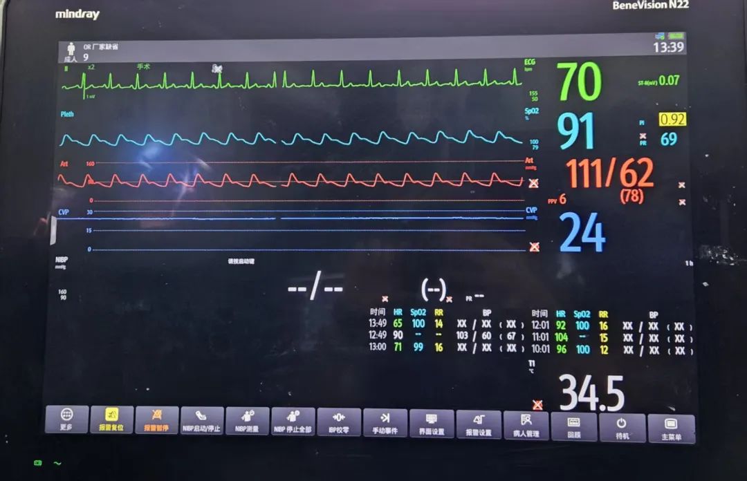
Post-anesthesia pulse oxygen saturation (SpO2) with pure oxygen: 91%

Surgical Review
Puncture performed with a puncture needle
Transvalvular device delivered into the left atrium
Sheath tube delivered into the left atrium
Clip opened in the left atrium
Mitral leaflets captured and clipped
Clipping position and effect confirmed
Clipping position confirmed via 3D imaging
Mitral regurgitation significantly improved
Mitral regurgitation status under 3D view
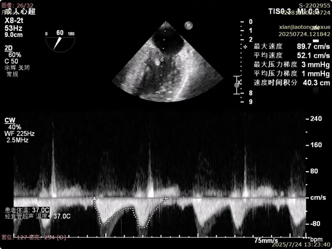
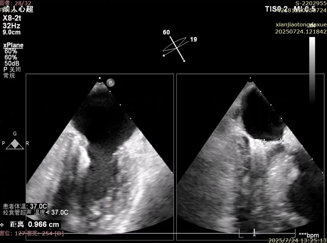
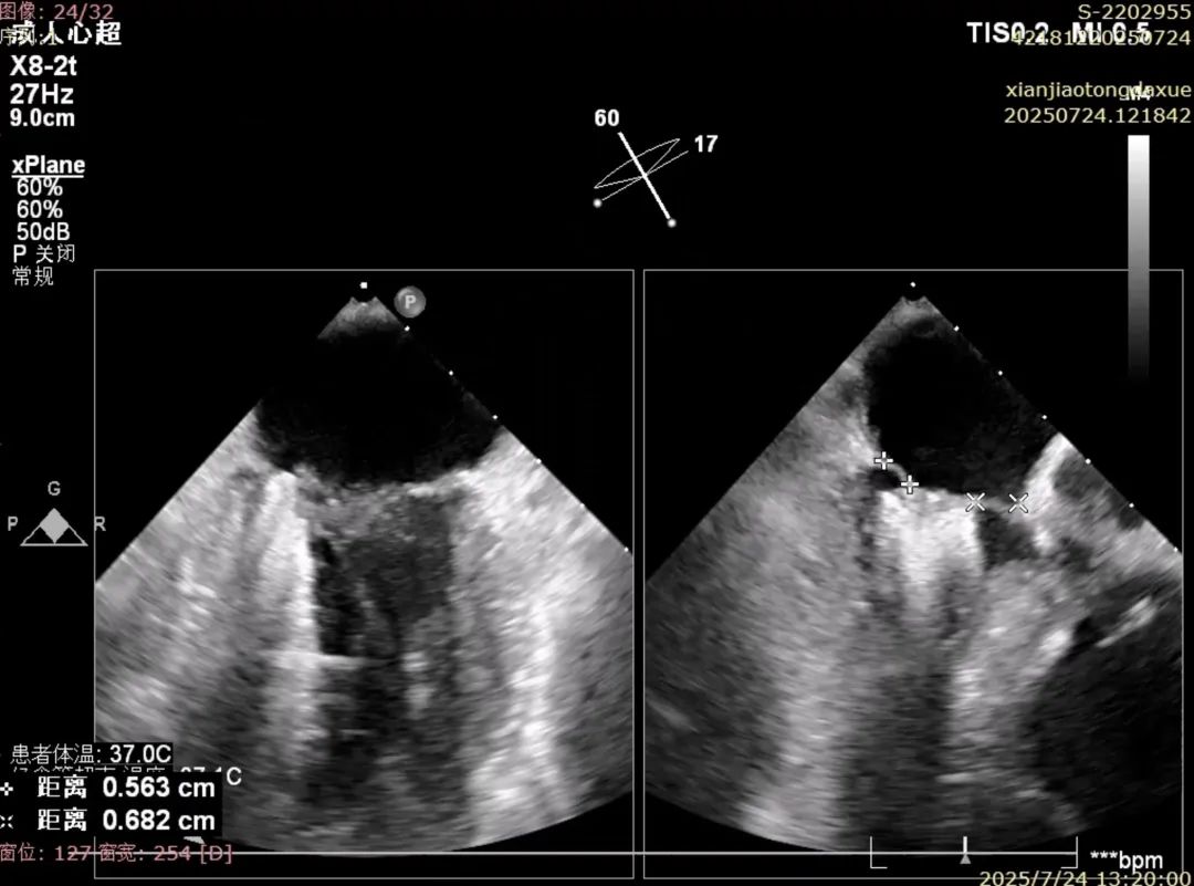
Postoperative clipping data confirmed, meeting expected outcomes
The success of this procedure not only demonstrates the adaptability and operational performance of domestic medical devices in complex lesions but also serves as recognition of the long-standing efforts of the team at the First Affiliated Hospital of Xi'an Jiaotong University in the field of minimally invasive valve intervention.
The First Affiliated Hospital of Xi'an Jiaotong University will continue to focus on the introduction and innovation of cutting-edge technologies for valvular heart disease treatment, promote the standardized and systematic development of minimally invasive cardiac therapy, and bring high-quality treatment and hope to more patients.


TEL: 021-6219 9996
ADD:Building 14, Gemdale Weixin Technology Park, 1288 Zhongchun Road, Minhang District, Shanghai